free Protein S - Protein S – a standard thrombophilia diagnostic parameter
What is thrombophilia?
Thrombophilia is a medical condition characterized by an increased risk to develop a thrombotic event. In most cases this is venous thromboembolism (VTE), such as deep vein thrombosis (DVT) or pulmonary embolism (PE). Also, the condition can lead to premature birth and other complications during pregnancy. Thombophilia diagnostics includes the determination of the underlying root cause for recurrent thrombotic events.
free Protein S deficiency as thrombophilia risk factor
Protein S is a Vitamin K depended protein, synthesized in the liver. It acts in the body as a natural anticoagulant. As co-factor of activated Protein C, it triggers the inactivation of the clotting factors Va and VIIIa.
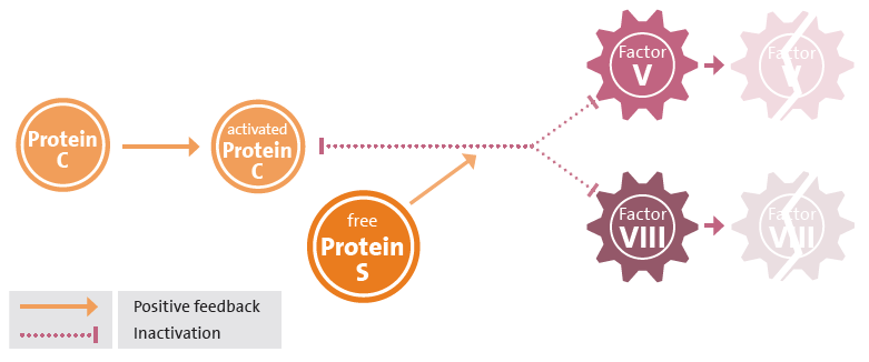
Dysfunctional or reduced Protein S can be the reason of an imbalanced coagulation system that results in thrombophilia. Approximately two thirds of Protein S is bound to C4 Binding Protein (C4BP). Only non-bound free Protein S has functional activity. The free Protein S level is expressed in %, with 100% corresponding to 1 IU/ml of the International Standard NIBSC 03/228.1
Forms of Protein S deficiencies
There are two forms of Proteins S deficiency, it can be aquired or hereditary. The hereditary form of protein S deficiency is caused by a mutation in a gene called PROS1. This condition is inherited in an autosomal dominant manner. The aquired form of Protein S deficiency originate either in a chronic or accute diseases or in medication.1
Causes of aquired Protein S deficiency
- Vitamin K-antagonist therapy
- Chronic infections
- Severe hepatic disease
- Nephritic syndrome
- Disseminated intravascular coagulation (DIC)
- Oral contraceptives
- Pregnancy
- Sickle cell anemia
Hereditary forms

- Heterozygous mutations cause a mild Protein S deficiency. However, almost half of all individuals with heterozygous Protein S deficiency will become symptomatic before the age of 55.1
- Homozygous mutations are very rare and cause a severe Protein S deficiency. It presents in neonates soon after birth and has a characteristic presentation of purpura fulminans.
Types of hereditary Protein S deficiencies
| Type I: | Decreased levels of total Protein S and free Protein S |
|---|---|
| Type II: | Decreased activity, but normal levels of total Protein S and free Protein S |
| Type III: | Normal level of total Protein S, but decreased level of free Protein S 1 |
Advantages of an immunologic free Protein S assay
Approx. 95% of patients have type I or III. For this reason a free Protein S assay has a higher predictive value than a total Protein S assay.2 Functional assays depend on various factors that influence coagulation. Hence the specificity of a functional assay is moderate with 40-70%.3 A free Protein S antigen assay utilizes specific free Protein S antibodies, that are not influenced by coagulation interferences.
Ordering information and specification
| REF | Format | Unit/Size |
|---|---|---|
|
|
||
| 36201 | Complete kit | 2 x 2,5 ml |
- Liquid, latex-enhanced immunoassay
with turbidimetric detection - Proven specificity for free Protein S
- Traceability to International Standard NIBSC 03/228
- Quality Control with HEMOSTAT Calibrator
and Control Plasma - Kit size also suitable for low-throughput
- Excellent open vial stability of 8 weeks (2-8°C)
- Regulatory status: CE IVD
| REF | Format | Unit/Size |
|---|---|---|
HEMOSTAT CALIBRATOR |
||
| 35500 | Calibrator | 4 x 1 ml |
- Lyophilized human plasma
| REF | Format | Unit/Size |
|---|---|---|
HEMOSTAT CONTROL PLASMA |
||
| 35001 | Normal | 6 x 1 ml |
| 35002 | Abnormal | 6 x 1 ml |
- Lyophilized human plasma
- Convenient in use through freeze-thawing capability
References
1. Gupta et al., Protein S Deficiency StatPearls Publishing LLC, 2020
2. Persson K.E. et al., Diagnosing protein S deficiency: analytical considerations. Clin. Lab. 49:103-110, 2003
3. R. Marlar & J. Gausman, Protein S abnormalities: a diagnostic nightmare Am. J. Hematol. 86:418-421, 2011

